MiCD Clinical Techniques
Discover BioLogically Beautiful Restorations
MiCD GN aims to provide educational resources to help dentists embrace the MiCD concept and treatment protocols in routine clinical practice.


Case prepared by Dr. Sushil Koirala (NP)
Missing & Peg-shaped Laterals
A middle aged (45 years) patient requested modification of a peg shaped lateral incisor. Upon examination a missing lateral incisor (#12), peg shaped lateral incisor (#22) and hypocalcified spots in the upper central inciors with broken incisal edges were noted. The shade distribution and surface texture of anterior teeth were not uniform. After his dental problems and cosmetic solutions were explained, the patient requested for a cost-effective treatment option. Therefore the case was completed with direct bonding and cosmetic contouring. The patient preferred to maintain the white spots on the central incisors. The main clinical challenge in this case was to obtain uniform surface texture and shade of all maxillary anterior teeth.


Figure 1
Peg shaped tooth #22, missing tooth #12 with uneven incisal edges of teeth #11 & #21.
Figure 2
Restored tooth #22 with direct cosmetic veneer and cosmetic contouring on #11, #21, #12.
Figure 3.

Figure 4
Before treatment.


Figure 5
Roughen surface of tooth #22 with a Diamond Point #102R.
Figure 6
Protect the adjacent teeth with a metal strip and etch with phosphoric acid.

Figure 7
The etched enamel surface with a frosted appearance.


Figure 8
Place a plastic strip and apply the bonding agent.
Figure 9
Light-cure the binding agent for 10sec.

Figure 10
Inject the flowable resin to create the frame.


Figure 11
Build-up the dentin (#A3) on the frame at distal margin.
Figure 12
Build-up the final dentin shape with (#A3).

Figure 13
Build-up the enamel layer (#INC) incrementally.


Figure 14
Smoothen the enamel surface with a brush and light-cure.
Figure 15
Finish the restored surface with Diamond Point #F104R.

Figure 16
Fine finish the labial surface with Diamond Point #SF215.
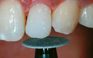

Figure 17
Contour the incisal edge of tooth #22 with Super-Snap black disk.
Figure 18
After restoration, note the colour, gingival shape and texture of tooth #21.

Figure 19
Finally super polish the surface with Super-Snap red disk.


Figure 20
Polish interproximal area with Super-Snap polystrips.
Figure 21
Use a diamond polishing paste with a Buff disk to achieve the final gloss.

Figure 22
Mark the area for contouring on Teeth #13, #11 & #21.


Figure 23
Contour uneven incisal edges with Diamond Point #102R and maintain the white spots.
Figure 24
Finish and polish teeth with OneGloss Midi Point and super polish with Buff disk.

Figure 25
Tooth #13 is transformed to mimic a lateral incisor.


Figure 26
Teeth #11, #21, #22 see after contouring and restoration.
Figure 27
Upper 4 anterior teeth seen after cosmetic contouring & restoration.
Preserving Health, Enhancing Smiles
Minimally Invasive Cosmetic Dentistry
(MiCD) is a holistic patient-centric treatment approach
